Cephalometric Imaging
Cephalometric Imaging
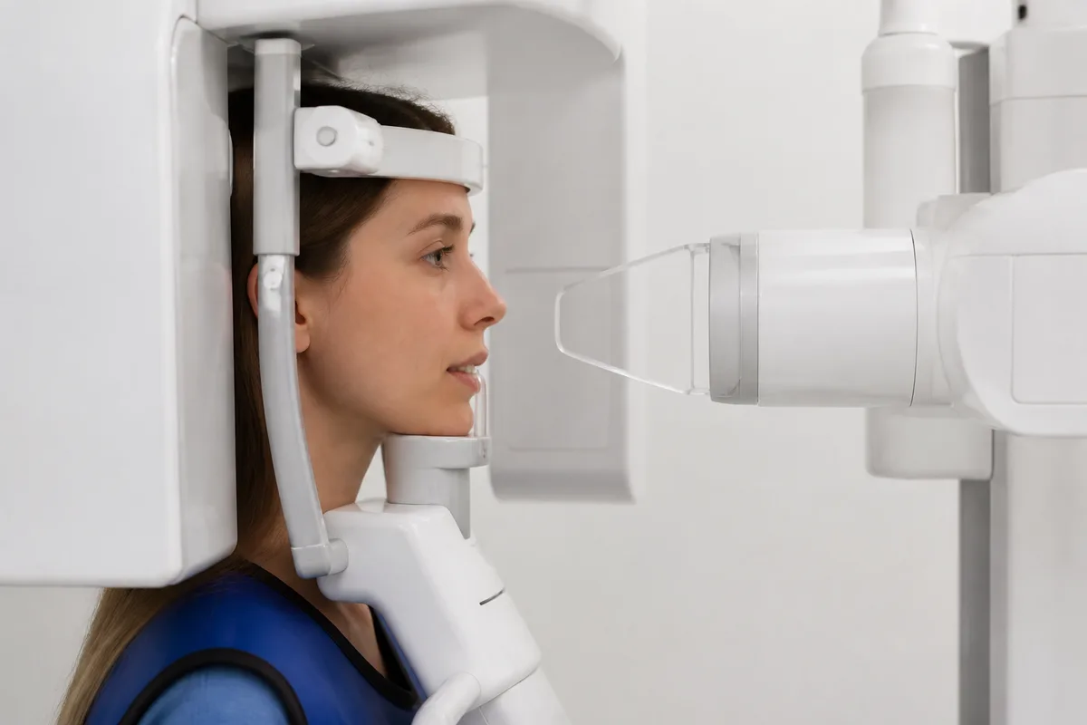
Cephalometric imaging is a side-profile skeletal X-ray that captures the relationship between the jaw, the facial bones, and the bite. It gives a clear view of structures that a front-facing photo or scan cannot show, helping guide decisions about growth patterns and treatment timing.
What It Helps Evaluate
- Jaw Alignment — shows how the upper and lower jaw relate to each other and to the skull.
- Growth Patterns — useful for tracking how the jaw is developing in younger patients.
- Bite Relationships — supports diagnosis of overbite, underbite, and other alignment concerns.
Part of a Complete Diagnostic Picture
Cephalometric images are often reviewed alongside digital panoramic X-rays and 3D treatment planning to build a full picture before any appliance or bracket system is recommended.
Who Reviews Your Images
At KHL Orthodontics, cephalometric images are reviewed by Dr. Kristine M. Hyon, DMD, MS, who completed her orthodontic residency and a Master of Science in Oral Biology at UMDNJ, as part of her evaluation of jaw alignment and bite development.
This content is for general information only and isn't dental or medical advice. Talk with a qualified provider about your own situation. If you have a medical emergency, call 911.