Digital Panoramic X-Ray
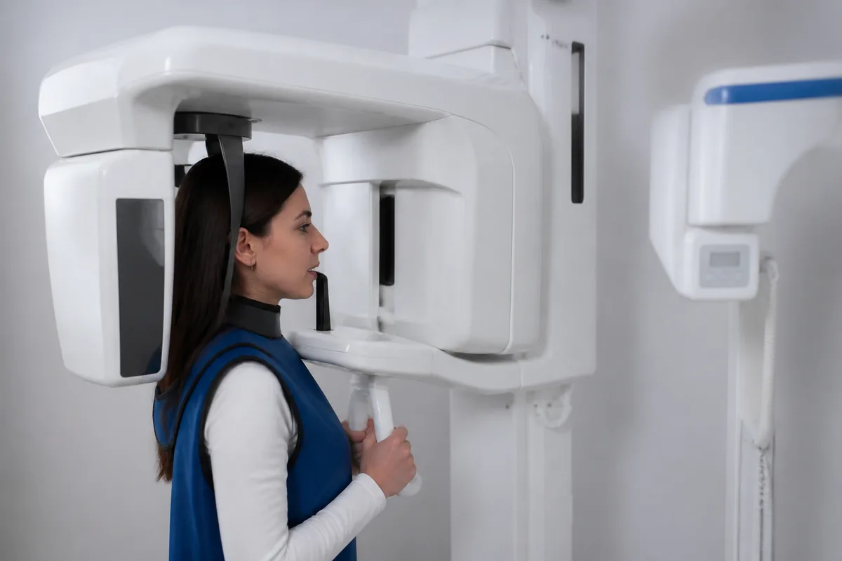
Digital Panoramic X-Ray
A digital panoramic X-ray captures a single wide image that shows the upper and lower jaw, the roots beneath the gumline, and the surrounding bone in one pass. Orthodontists use this broad view to check how everything is positioned before mapping out a treatment plan, and to watch how growth and movement progress over time.
Why This Imaging Matters
- Full-Jaw View — Shows both arches, roots, and supporting bone in a single scan.
- Low Radiation — Delivers detailed imaging while keeping exposure low.
- Foundation for Planning — Gives a clear starting point before appliances or aligners are introduced.
Working With Other Diagnostic Tools
Panoramic imaging is often paired with cephalometric imaging for a side-profile skeletal view, and with 3D treatment planning to preview how movement will unfold. Together, these tools give a fuller picture of jaw structure and alignment.
Who Reviews the Images
At KHL Orthodontics LLC, Dr. Kristine M. Hyon, DMD, MS, reviews imaging as part of building an individualized treatment plan for each patient.
This content is for general information only and isn't dental or medical advice. Talk with a qualified provider about your own situation. If you have a medical emergency, call 911.